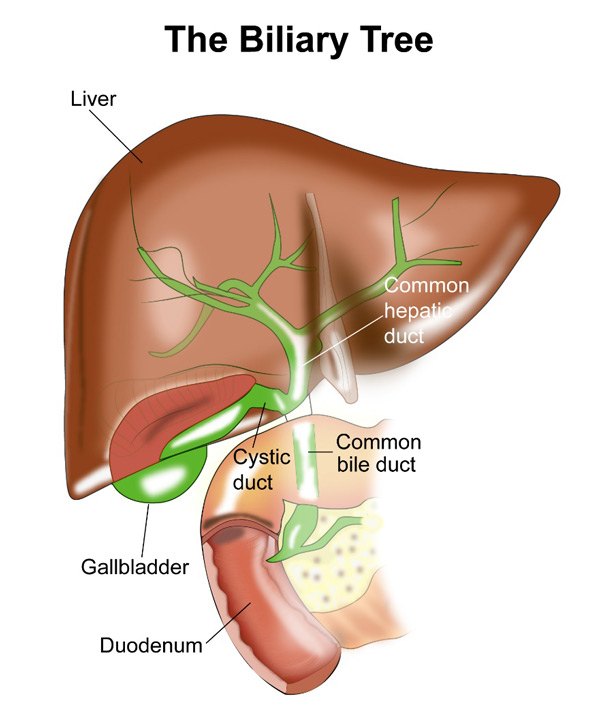
Gallbladder is a small pouch or sac present under the liver which stores and concentrates bile. Bile is a liquid produced by the liver and is used to digest food such as fats. The bile in the gallbladder flows into the common bile duct which opens into a part of the small intestine called duodenum. At the time of digestion of food, the gall contracts and the bile is released into the duodenum.
A cancer that develops in the gallbladder is called gallbladder cancer. This cancer is usually an adenocarcinoma.

Gall Stones
People who have gall stones have a very small increase in risk of developing gallbladder cancer. Having gall stones is very common and only about 1 in 200 people develop gallbladder cancer.
Age
The risk of developing gallbladder cancer increases with age and usually happens in people over the age of 70
Smoking
Smoking increases the risk of developing gallbladder cancer.
Chronic Infection
There is an increased risk of developing gallbladder cancer in people who are carriers of salmonella infection. Carriers are people who have this infection but do not have any symptoms associated with it. Salmonella infection causes typhoid fever. North India has a high rate of gallbladder cancer and this infection may play a part in these high rates.
Obesity
Being obese increases the risk of developing gallbladder cancer as well as many other cancers. About 10%-20% of gallbladder cancer can be related to obesity.
Gall bladder Polyps
Polyps are benign growths that can develop in gallbladder. People known to have gallbladder polyps have an increased risk of developing gallbladder cancer.
Family History
Having a close relative like parent, brother or sister with gallbladder cancer increases the risk.
Gallbladder cancer may or may not produce any symptoms in the early stages. The following symptoms may be produced by gallbladder cancer.
Jaundice
Jaundice is yellowish discolouration of the eyes and the skin. This symptom can be present in a number of medical conditions including cancer and gallbladder cancer. The jaundice is usually painless and gradually increases in severity. A doctor should be consulted when jaundice is seen or suspected in a patient.
Pain
Patients with gallbladder cancer can have a pain in the abdomen, particularly in the right side of abdomen. This is usually a dull aching type of pain. Sometimes the pain can be more severe and sharp.
Other symptoms
In patients with advanced disease, symptoms such as reduction in weight and appetite as well as weakness can be present. Some patients may notice swelling of the abdomen or feeling of a lump or mass in the abdomen or nausea and vomiting.
When a Gallbladder cancer is suspected or diagnosed, the following investigations are done to complete the diagnosis and staging of the cancer.
US Abdomen
An ultrasound scan of the abdomen particularly the biliary tract is done first in patients presenting with jaundice. This scan will look for evidence of obstruction of the bile duct. Obstruction of the bile duct can also happen for reasons other than cancer such as gallstones etc. If the ultrasound shows a suspected cancer, then further tests are needed.
CT Scan
A CT scan of the abdomen is an important test in the diagnosis of gallbladder cancer. It helps identify a tumour mass present within the gallbladder and can aid in the biopsy of the abnormal mass.
ERCP
An ERCP is endoscopic retrograde cholanio pancreatography, a procedure where a gastroenterologist passes an endoscope (narrow tube passed into the stomach) into the duodenum and from there gains access into the opening of the common bile duct through which the bile and the pancreatic enzymes drain into the duodenum. Once access is gained into this duct, the cause for the blockage of the duct is found by injecting a dye and taking x-ray pictures. If cancer is found or suspected, a biopsy can be taken from the site. This procedure is done under sedation. A stent may be placed sometimes to keep the duct open. This helps relieve the jaundice.
MRI
An MRI scan is sometimes done to investigate gallbladder cancer. An MRCP is magnetic resonance cholangio pancreatography can give the same information as an ERCP about possible causes of bile duct blockage but a biopsy cannot be done or a stent cannot be placed as this is a non invasive test.
EUS
An EUS is endoscopic ultrasound where an endoscope is inserted into the gullet and stomach. The endoscope has a small ultrasound probe attached to it which helps get an ultrasound image from the inside. This test helps in getting clearer images of small masses or lymph nodes present in the area. The test also guides the doctor to take biopsies accurately.
Staging Laparoscopy
A staging laparoscopy is a test where a laparoscopic procedure is done to look for presence of incurable disease or for a biopsy. A laparoscopic procedure involves placing small cuts on the abdomen and inserting long tubes into the abdomen through these cuts. These tubes have a light and camera source attached to them and the doctor can have a look inside the abdomen with them.
PET-CT
A PET-CT scan is not routinely recommended as a standard investigation for gallbladder cancer but can sometimes be used in addition to standard CT discussed above.
Biopsy
A biopsy is not done prior to surgery if all investigations suggest definite gallbladder cancer. It is recommended though in advanced tumours when other forms of treatment such as chemotherapy or radiotherapy are planned prior to surgery. A biopsy can be done with the help of ERCP or EUS or CT guided. Some clinicians advocate a biopsy prior to any treatment.
The stage of a cancer is a term used to describe the size and location of the cancer in the body.
Knowing the stage of the cancer helps the doctors to decide on the most appropriate treatment. Gallbladder cancer is staged based on the TNM staging system or the number system.
Staging with either system is based on the extent of the tumour in the gallbladder, the spread of the cancer into the lymph nodes, and spread of cancer into other parts of the body.
TNM stands for Tumour, Node and Metastases. T stands for tumour and in gallbladder cancer represents depth of spread into the wall of the gallbladder. N stands for nodes and spread of cancer into nodes. M stands for metastases and spread of cancer to distant sites in the body.
T stage
| T1 | Tumour invades into the lamina propria or muscular layer |
| T1a | Tumour invades into lamina propria |
| T1b | Tumour invades into muscular layer |
| T2 | Tumour invades tissue around the muscle; no extension beyond serosa or into liver |
| T3 | Tumour perforates the serosa and/or directly invades the liver and/or one other adjacent organ or structure, such as the stomach, duodenum, colon, pancreas, omentum, or extrahepatic bile ducts |
| T4 | Tumour invades main portal vein or hepatic artery or invades two or more organs or structures outside the liver |
N Stage
| N0 | No regional lymph node involvement |
| N1 | Spread of cancer to lymph nodes along the cystic duct, common bile duct, hepatic artery, and/or portal vein |
| N2 | Spread to periaortic, pericaval, superior mesenteric artery, and/or celiac artery lymph nodes |
M Stage
| M0 | No distant metastasis |
| M1 | Distant metastasis |
Treatment of Gallbladder cancer involves many options including Surgery, Chemotherapy and Radiotherapy. Which strategy to select depends on the age of patient, fitness of the patient, stage of cancer at diagnosis and the individual preferences of the patient.
Surgery for Gallbladder Cancer
Surgery is the most important and the only curative option in the treatment of this type of cancer. Surgery will be an option in early and some locally advanced cancers. The type of surgery selected will depend on the extent of disease on staging investigations. For very early cancers, simple removal of gallbladder (cholecystectomy) is enough. For more advanced cancers, the gallbladder with a portion of normal liver around it is removed at operation. This is known as extended cholecystectomy. These surgeries may involve removal of the bile duct and usually involves removal of surrounding lymph nodes.
In patients with very advanced disease where curative surgery is not possible, surgery is sometimes done to improve symptoms such as jaundice or pain.
Adjuvant Treatment for Gallbladder Cancer
Adjuvant treatment is where additional treatments are given after the main curative treatment of cancer is completed. This type of treatment aims to increases the chances of cure. In this cancer, adjuvant treatments that are offered include Chemotherapy alone or Chemoradiotherapy which is a combination of Chemotherapy and Radiotherapy. In patients not suitable for Chemotherapy, adjuvant Radiotherapy alone can also be used. Which option to use depends on the pathology report after the surgery and the condition of the patient. When Chemotherapy alone is used, it is given for up to 6 months.
Chemotherapy for Gallbladder Cancer
Chemotherapy is used for gallbladder cancer in different settings. It can be used as adjuvant treatment after surgery as described above. It is used commonly in advanced or metastatic cancer when surgery is not possible. Here chemotherapy is used using one drug or a combination of drugs. The choice depends on the fitness of the patient. Common drugs used here include Gemcitabine, Capecitabine, 5-Fluorouracil, Cisplatin, Carboplatin or Oxaliplatin. In patients who are fit, a combination of two drugs are given for up to 6 months.
Radiotherapy in Gallbladder Cancer
Radiotherapy is used in gallbladder cancer either alone or in combination with Chemotherapy as described above in the adjuvant setting. Radiotherapy is also used to treat symptoms such as pain in advanced disease.
Supportive Measures
In patients with Gallbladder cancer, particularly advanced disease, jaundice is a dominant symptom and relieving the jaundice is a important option of treatment. If there is a blockage of a bile duct, a stent can be inserted endoscopically which relieves the jaundice. If stent is not possible other options such as bypass surgery are done.


