

Rectum is a part of the large intestine present in the lower part of the tummy called the pelvis.
A cancer that develops in the rectum is called as Rectum cancer. This cancer is usually an adenocarcinoma.
The function of the rectum is to store the faecal matter after digestion of the food before it is expelled outside through the anus.

Rectal cancer is cancer that starts or originates in the rectum. Cancer that develops in the rectum starts in the innermost lining of the rectum called the mucosa. Cancer usually originates from the gland-like cells present in the mucosa and is called an adenocarcinoma. Other types of rectal cancer include squamous cell carcinoma, but this is very rare.
Rectal cancer can spread from the mucosal layer into other layers of the rectum and then to the outside of it. This type of spread is called the direct spread. It can also spread through the lymphatics into the lymph nodes around the rectum and to other parts of the body. Cancer also spreads through the bloodstream to other parts of the body such as the liver and lungs.
Rectal Cancer can present with a variety of symptoms and these are listed below
Change in bowel habit
Change in bowel habit is a common symptom seen in rectal cancer. This change can be in the form of unexplained diarrhoea or constipation or alternating diarrhoea and constipation. These symptoms if present for more than three weeks, warrants investigations.
Blood in the motions
Passing blood from the anus or blood in the motions can be a symptom related to colon or rectal cancer. Most times, passing blood in the motions is not due to cancer and due to piles or other causes but it is important to see a doctor to have it checked out.
Abdominal Pain
Abdominal or tummy pain can be a symptom of bowel cancer. Again, there will be many causes for this symptom and tests need to be done to look for a cause if the symptom persists.
Other Symptoms
Other symptoms such as weight loss, tiredness, anaemia, distension of abdomen, vomiting can all be associated with colon cancer.
Age
Like all cancers which are more common in older age, the risk of rectal cancer is more in patients in the older age group with majority of cancers occurring in those aged over 60.
Genetic Factors
5% of rectal cancers are passed on in families due to an abnormal gene. Genetic link to rectal cancer is suspected if more than one close family member is affected with cancer at any age or one close relative had cancer diagnosed before the age of 45. A close family member is a first degree relative who could be a parent, child, brother or sister.
In the 5% of rectal cancers that are due to faulty genes inherited from the family, the two conditions commonly seen are Familial adenomatous polyposis(FAP) and Hereditary non polyposis colon cancer(HNPCC). FAP leads to number of polyps in the rectum at a young age which over time can turn into cancer. In those with confirmed FAP, surgery is recommended to remove the colon and rectum to reduce the risk of these polyps turning into cancer.
HNPCC is another condition associated with colorectal cancer as well as other cancers such as pancreas, bladder, uterus and ovarian cancers.
Obesity and Physical Activity
Being obese is a risk factor for rectal cancer. Also, lack of physical activity seems to increase the risk.
Diet
Diet rich in red and processed meat increases the risk of rectal cancer. Rich diet of red and processed meat involves eating these foods on most days of a week. Consumption of red meat once or twice a week doesn’t increase the risk.
A diet rich in fruit and vegetables reduces the risk of getting colon or rectal cancer.
Alcohol
Alcohol consumption increases the risk of developing rectal cancer and the risk increases proportionally to the amount of alcohol taken.
Inflammatory Bowel Disease
Patients with a long term history of inflammatory bowel disease such as Crohn’s disease and Ulcerative Colitis have a higher risk of developing colon and rectal cancer. This risk is increased after 10-20 years from diagnosis of inflammatory bowel disease.
Abdominal Radiotherapy
Adults who received lower abdominal radiotherapy as a child for cancer, have a higher risk of developing a rectal cancer in adulthood.
If rectal cancer is suspected, the following investigations are usually done. Not all investigations are needed in every case.
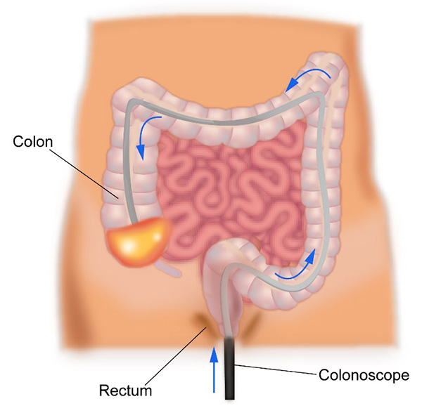
Colonoscopy
A colonoscopy is an endoscopic test where a thin tube with a light and a camera at its end is inserted into the anus and passed up into the rectum and colon. The doctor doing the test will be able to look at the inside of the colon and rectum on a screen and will be able to identify any abnormalities present. The whole of the colon and rectum can be seen through this procedure.
It is an outpatient procedure and is done under mild sedation to reduce the discomfort. If the doctor finds anything abnormal, a sample of the tissue can be taken(biopsy) for tests.
Also, sometimes small abnormal areas like polyps can be removed completely with this procedure. Before the colonoscopy, the patient is given medicines to take the day before to help empty the colon and rectum.
Flexible sigmoidoscopy
This test uses a similar tube as a colonoscope but is used to examine only the lower part of the colon and rectum. This is done as an outpatient procedure and usually does not need any sedation. An enema is given prior to the test to empty the rectum and lower colon.
Virtual Colonoscopy or CT Colonography
This test involves looking inside the colon with the help of a CT scan. The patient is given medicines the day before to empty the colon and advised regarding a specific diet for a few days before the scan. A CT scan is done to get detailed images of the colon. Any abnormalities present in the colon can be seen. The drawback of this test is that a biopsy cannot be taken, and a colonoscopy needs to be done again to get a biopsy.
CT scan
Once a colon cancer is diagnosed on colonoscopy and biopsy, a contrast enhanced CT scan is done of the chest abdomen and pelvis to stage the cancer. This scan is used to look for spread of cancer from its point of origin to other parts of the body.
PET-CT Scan
A PET-CT scan is a PET scan combined with a CT scan. It involves injecting a special radioactive dye into the vein prior to the scan. This special dye will enable to detect cancer better than normal CT scan in certain circumstances.
In rectal cancer, PET-CT is not recommended for routine staging but is recommended in settings when surgery is being considered to the liver or other organs or when standard CT scan is unable to determine clearly the presence or absence of cancer.
MRI Scan
An MRI scan is routinely used to stage or diagnose rectal cancer. It is a very accurate test to identify the spread of rectal cancer locally and to assess the liver specifically when abnormalities present in the liver are suspicious for cancer or when liver surgery is being contemplated. This is a very useful test in this type of cancer to give an accurate local staging and help in decision regarding the best treatment.
Endorectal Ultrasound
This is an ultrasound scan of the rectum done to stage the rectal cancer accurately. It can be done instead of an MRI. It helps define the spread of the cancer through the layers of the rectum. The procedure is done by inserting an ultrasound probe into the rectum.
Biopsy
A biopsy of the mass or tumour in the rectum is taken to confirm the diagnosis of rectal cancer. The biopsy is done with the help of the above tests. Molecular tests of KRAS, NRAS and BRAF are done on the biopsy sample particularly in patients with stage 4 cancer as this helps in the decision process.
The stage of a cancer is a term used to describe the size and location of the cancer in the body.
Knowing the stage of the cancer helps the doctors to decide on the most appropriate treatment. Rectal cancer is staged based on the TNM staging system or the number system.
Staging with either system is based on the extent of the tumour in the rectum, the spread of the cancer in the rectum and into the lymph nodes and spread of cancer into other parts of the body.
TNM stands for tumour, node and metastases. T stands for tumour and in colon cancer represents depth of spread into the wall of the rectum. N stands for nodes and spread of cancer into lymph nodes around the rectum. M stands for metastases and spread of cancer to distant areas in the body.
Number staging system
Based on the TNM stage of the cancer, rectal cancer can be divided into stages 1 to 4 as below. Duke’s staging is another classification of rectal cancer. Duke’s staging is from A to D.
Stage 1
In stage 1, the cancer is limited to the rectum and involves the part of the rectum called the submucosa and muscularis layers. There is no involvement of the lymph nodes.
Stage 2
In stage 2, the cancer involves into the areas outside the colon or into surrounding organs without any involvement of the lymph nodes.
Stage 3
In stage 3 cancer, the tumour can involve any layer of the rectum but there is definite involvement of the lymph nodes.
Stage 4
In stage 4, the cancer has spread to different parts of the body such as liver, lungs etc.
The treatment of rectal cancer is dependent on the stage of cancer at diagnosis. The staging process will divide the rectal cancer into non metastatic, where disease is confined to one area of the rectum and lymph nodes with no evidence of spread to distant sites in the body and metastatic cancer where cancer has spread from its site of origin to distant parts of the body.
Treatment also depends on the symptoms present at diagnosis irrespective of the cancer being metastatic or non-metastatic.
By and large, rectal cancer that does not show distant spread is treated with surgical removal, where the rectum with the cancer and surrounding lymph nodes are removed. Details of surgery are given below. Except in very early rectal cancer, a combination of radiotherapy and chemotherapy lasting 5-6 weeks is given to the cancerous area prior to surgery. This treatment is called as neo-adjuvant chemoradiotherapy. The purpose of this treatment is to reduce local recurrence of rectal cancer after surgery. Following surgery, chemotherapy is advised based on the pathology results from the surgery. Chemotherapy given in this setting is called adjuvant chemotherapy where the purpose of it is to maximise the chances of a cure.
In a few instances, when neoadjuvant chemoradiotherapy is not done prior to surgery, adjuvant chemoradiotherapy (after surgery) can be considered but this is not very common.
In patients with metastatic cancer, chemotherapy is usually the first treatment of choice. Here chemotherapy can be given on its own or along with biological agents, the details of which are discussed below.
In patients with metastatic disease but with symptoms suggesting of obstruction in the rectum, an operation with insertion of a stoma or a stent insertion into the rectum is done to improve the symptoms of obstruction. Surgical removal is done first followed by chemotherapy. Patients with limited metastatic disease confined to parts of liver or lung are treated with surgery to remove the cancer in the rectum and at metastatic sites.
Radiotherapy is used in different settings in rectal cancer and these are listed before
Neoadjuvant chemoradiotherapy
A treatment is called as neoadjuvant when it is given prior to a definitive form of treatment. In rectal cancer with no distant spread, the definitive treatment is surgical removal.
Radiotherapy along with chemotherapy given prior to this treatment is called as neoadjuvant chemoradiotherapy. Here, the main treatment is radiotherapy with the chemo given along with it to make the radiotherapy more effective. The treatment is advised for most rectal cancers except for very early ones.
Radiotherapy is given to the lower part of the tummy called the pelvis where the rectum and local lymph nodes are present. It is done once a day, 5 days a week for 5-6 weeks. Chemotherapy can be given as tablets or through a drip at the same time as radiotherapy over the 5-6 week period. The tablet chemotherapy that is used is called as Capecitabine and is used twice a day. If a drip or intravenous version is used, this drug is called as 5-Fluorouracil. Different techniques of radiotherapy can be used including 3D conformal radiotherapy, Intensity modulated radiotherapy (IMRT), or arc based therapy (VMAT, Rapid arc).
Following completion of this treatment, a gap of 8-12 weeks is given before surgery is done to remove the tumour.
In instances where neoadjuvant treatment is not given prior to surgery, it can be given after surgery is done and this is known as adjuvant chemoradiotherapy. The purpose of the treatment remains the same, only that surgery is done first. Generally neoadjuvant chemoradiotherapy is preferred over adjuvant chemoradiotherapy.
Side effects of chemoradiotherapy include tiredness, loose motions, redness of the skin and soreness of the skin, pain and discomfort while passing motions, risk of infection, discomfort or a burning sensation while passing urine and passing of blood in the motions. These side effects are present from about the third week of treatment and can last up to 4-6 weeks after completion of treatment.
Preop short course Radiotherapy
This is also neoadjuvant treatment as above but is not given along with chemotherapy. It is radiotherapy alone and is given only for one week just before surgery. It is used as a treatment in early rectal cancer and is practiced commonly in Europe but not so much in India.
Palliative Radiotherapy
Here, radiotherapy is used to control symptoms produced by rectal cancer when the cancer cannot be cured anymore. The aim of treatment here is to control symptoms such as pain, bleeding and blockage in the rectum. The treatment duration can be variable ranging from 1-25 treatments depending on where the treatment needs to be given and the symptoms that need controlling.
Surgical removal is the most important component of curative treatment of rectal cancer.
There are different types of operations done to remove rectal cancers and the choice of operation depends on the stage of cancer, its location in the rectum, local surgical expertise and fitness of the patient. The different options are listed below.
Local Excision
This is a procedure done in very early stage rectal cancers. Here the surgeon inserts an endoscope into the rectum through the anus and removes the cancer. Following the removal, the cancer is looked at by the pathologist for completeness of removal. If removal of cancer is incomplete or the stage of cancer is more advanced than expected, more surgery may be needed. If removal is complete, no further treatment is needed. Different types of local excision are present including trans anal excision (TAE) and transanal endoscopic microsurgery (TEM).
Total Mesorectal Excision
When a cancer operation is done, the aim of surgery is to remove the cancer with clear margins (cancer not present at the margin of resection). This is done by taking out a rim of normal tissue along with the cancer. In rectal cancer this is best achieved by doing a total mesorectal excision (TME).
Total mesorectal excision involves removal of the rectum containing the cancer in its entirety along with the fat tissue and the mesolectal tissue surrounding the rectum. The mesorectum contains the lymph glands which the cancer can spread into and removal of it removes the lymph glands too. Studies have shown this approach to provide the best chance of avoiding a local recurrence of cancer.
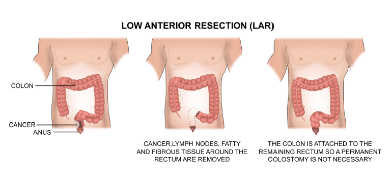
The type of TME that is done is dependent on the location of the cancer in the rectum. If the cancer is located higher up in the rectum, an anterior resection is done.
Anterior resection involves removing the diseased part of the rectum and joining the lower part of the colon to the remaining rectum. If the cancer is present in the middle part of the rectum, most of the rectum is removed and the lower part of the colon is attached to the anus. This is called a colo anal anastamosis. In some situations, when a very low anterior resection is needed, the surgeon may do a rectal reconstruction, where the colon is fashioned to function as a reservoir for the faeces. This helps in providing better function after the operation.
If a colo anal anastamoses is done, a temporary stoma may be needed to allow the bowel to heal. Sometimes, to allow the bowel to heal, an ostomy or stoma is done. The involves bringing the end of the cut bowel as an opening onto the abdomen. A bag is attached to the ostomy, into which faeces and fluid collects. This ostomy is a colostomy, if the colon is brought out or an ileostomy if the end of the small intestine is brought out. This procedure can be reversed at a later date.procedure can be reversed at a later date.
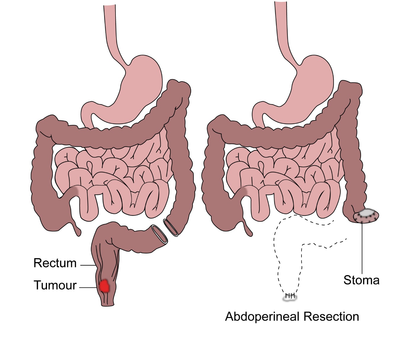
If the tumour is in the lower part of the rectum, an anastamosis may not be possible and the surgeon proceeds to do an abdomino perineal resection(APR), where the rectum and anus is removed and a permanent colostomy is formed.
The surgery can be done by the open method or minimally invasive method (laparoscopic).
Chemotherapy is an important component of treatment for Rectal cancer. Chemotherapy is given in a number of settings in this condition.
Adjuvant Chemotherapy
Adjuvant chemotherapy is given to patients after definitive surgical removal of the cancer. The purpose of adjuvant chemotherapy is to reduce the risk of recurrence of cancer after curative removal. Chemotherapy is definitely indicated in patients with Duke’s C cancer and some patients with Duke’s B cancer. The pros and cons of chemotherapy in this setting can be discussed with an Oncologist. The drugs commonly used are Fluorouracil, Capecitabine and Oxaliplatin. When Oxaliplatin is used, it is given in combination with Fluorouracil or Capecitabine. The duration of treatment can be 3-6 months and is given once every two or three weeks depending on the drugs selected.
Palliative Chemotherapy
Palliative chemotherapy is given when there is stage 4 cancer. The purpose of chemotherapy is to reduce the cancer and its symptoms and to prolong life.
Drugs used for stage 4 rectal cancer include chemotherapy drugs such as Fluorouracil, Capecitabine, Oxaliplatin, Irinotecan and Trifluridine-Tipiracil. Oxaliplatin is given in combination with Fluorouracil (FOLFOX) or Capecitabine (XELOX). Similarly, Irinotecan can be combined with Fluorouracil (FOLFIRI) or Capecitabine (XELIRI). These combination regimens are given once every two or three weeks depending on the regimen selected. The chemotherapy regimens can be combined with additional drugs such as antiangiogenic agents or anti EGFR antibodies, the details of which are listed below.
The duration of palliative chemotherapy ranges from 3-6 months for one drug or combination of treatment. The total duration of that particular regimen is dependent on response of the cancer to that treatment and the tolerance of the treatment by the patient.
Following completion of one regimen of treatment, further chemotherapy may be needed either soon after the first one or sometime later, depending on the status of the cancer at that time.
Chemotherapy prior to resection of metastatic disease
In some patients with stage 4 cancer, the disease is just confined to the liver or lung which may be amenable for surgical resection. In such a setting, chemotherapy is given for around three months prior to liver resection to maximise the chances of good control of disease. This chemotherapy can be restarted after surgery and continued for a total of six months.
Anti EGFR antibodies
Anti EGFR antibodies are drugs that target the EGFR receptor present on the cancer cell. This stops the cells from growing. Two drugs that are used are Cetuximab and Panitumumab. These are given alone or in combination with chemotherapy. Not all patients with colon cancer benefit from these drugs. At the time of biopsy, a special test (RAS testing) is done to determine whether a KRAS NRAS or BRAF mutation is present in the tumour or not. If KRAS, NAS or BRAF V600E mutation is present, these drugs will not be suitable. However, if KRAS, NRAS or BRAF mutation is not present (KRAS wild type), these drugs will be beneficial. Both Cetuximab and Panitumumab are given through the vein and are usually well tolerated. Common side effects of these drugs include skin rash, diarrhoea, sore mouth and tiredness.
Anti-Angiogenic Agents
Angiogenesis is growth and development of new blood vessels. Cancers need to develop new blood vessels to continue to grow. Anti-angiogenic drugs stop the development of new blood vessels and in turn stop the progression of cancer. The drugs available are Bevacizumab (Avastin), Aflibercept (Zaltrap) and Regorafenib (Stivarga). These drugs can be given on their own or in combination with chemotherapy. They are suitable to be given in all patients with stage 4 colon cancer and do not need any special testing. Common side effects of these drugs include high blood pressure, risk of bleeding, loss of protein in the urine, tiredness and risk of infection.
