

Diagnosis of Cancer
Once a cancer is suspected, tests are needed that will confirm or refute the suspicion. The following tests can be done to help in the diagnosis of cancer.
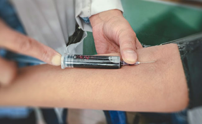
A variety of blood tests are done in the process diagnosis of cancer. Some blood tests are done for all cancers and some others are done to look for specific cancers. Specific blood tests include tumour markers which are proteins produced by the cancer and are present in the bloodstream and can be measured. Some blood tests are also done to see if the cancer is responding to treatment or not.
Common blood tests done include
- CBP (complete blood picture)
- Kidney function tests including electrolytes
- LFT (Liver function tests)
- ESR (Erythrocyte sedimentation rate)-done sometimes
- Calcium
- Clotting Profile (particularly before a biopsy)
Specific blood tests include
- PSA (Prostate specific antigen)- elevated in prostate cancer
- CEA (Carcinoembryogenic antigen)- elevated in colon and rectum cancers mainly
- CA153- elevated in breast cancer
- CA125- elevated in ovarian cancer
- CA19.9- elevated in Pancreatic cancers mainly
- AFP (Alfa feto protein)-elevated in Liver cancer and in testicular cancer
- B-HCG- elevated in testicular cancer
- Thyroglobulin- elevated in thyroid cancer
- Calcitonin- elevated in some thyroid cancers
- LDH (Lactate dehydrogenase)- elevated in lymphomas
- Serum Immnoglobulins- elevated in myeloma
- Serum Protein electrophoresis- tested in myeloma or plasma cell disorders
- Beta 2 Microglobulin- elevated in myeloma
It is important to note that having elevated levels of these tests doesn’t necessarily mean that there is a cancer in the body. These tests can be elevated in a benign conditions too. The tests are not very specific to the particular cancer. Some of these tests can be elevated in multiple cancers. Their importance is more in monitoring response to treatment once a diagnosis is already made and to look for recurrence of cancer.
With modern technology available, x-rays and scans form a major part of investigations that are done to help in the diagnosis of cancer. The following tests are commonly done.
X-rays
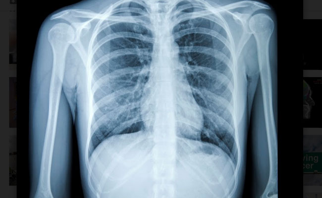
x-rays of the chest, breast (mammogram), abdomen, bones can be done to assess symptoms, to look for the presence of cancer and to assess response to treatment. However, x-rays are generally not as good as scans and any suspicion on x-rays will usually lead to a scan being done.
Ultrasound scan
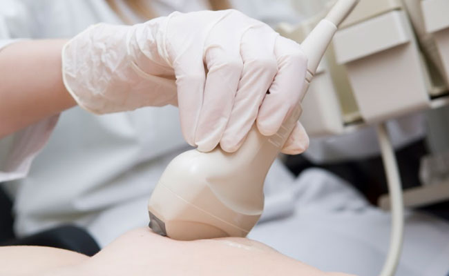
Ultrasound scans use sound waves to make images and are used commonly in diagnosis of breast and other cancers. These scans are cheap and harmless but may not be as good in detecting some cancers as other scans do.
CT Scan
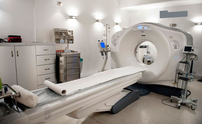
A CT scan is a scan that uses x-rays to give a three-dimensional image of the part of the body that is scanned. It is more accurate than an x-ray in identifying and staging cancer. A contrast enhanced scan is one where an injection is given into the vein prior to the scan and this gives better images. Oral contrast is given to the patient to drink when the abdomen is being scanned. CT scan takes only a few minutes to do. CT scan is also used to help guide the doctor when certain biopsies are done.
MRI Scan
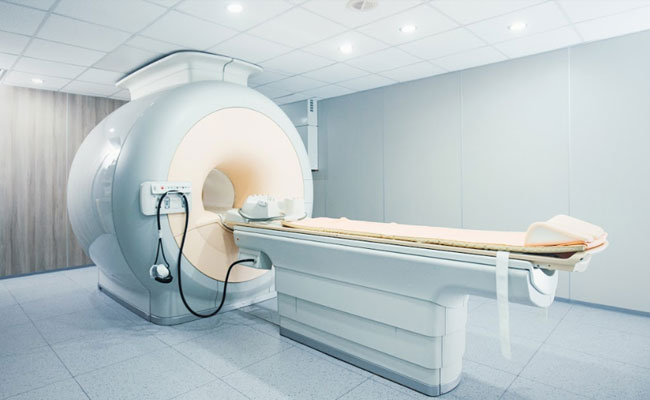
An MRI scan uses magnetic fields to generate images and is used very often to diagnose and stage cancer. This scan is done for some types of cancer. MRI produces better images than CT in areas such as brain, spine and pelvis (lower part of abdomen). Some patients find it difficult to have an MRI scan as they can feel claustrophobic while the scan is being done. The time taken for MRI scans is longer ranging from 20 mins to up to an hour. Contrast agent is injected during the scan to get better images. The scan is done as sequences and usually many sequences are done in one scan in cancer patients.
PET-CT Scan
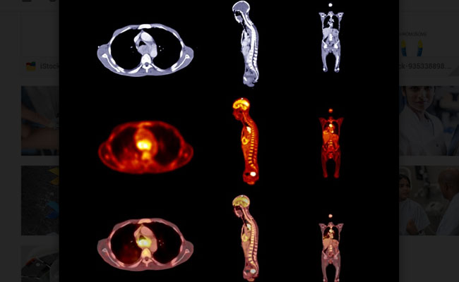
This type of scan differs from standard CT scan by having a functional element to the scan. The PET component of the scan is able to detect areas in the body where cells rapidly divide such as cancers, infection, inflammation etc. PET-CT is better than CT scan in staging certain cancers. The PET part of the scan is done by injecting a radiolabelled substance into the body first and then a scan is done. This substance is taken up by the dividing cells and shows up on the scan.
There are different types of PET-CT scan and the difference lies in the type of radiolabelled tracer used to detect the rapidly dividing cells. The standard PET-CT scan is an FDG PET which uses Fluorodeoxyglucose. The other tracers used include Choline C-11, 11 C Methionine, Fluorodeoxyglucose labelled Choline, Gallium 68 labelled somatostatin analogue scans used for neuroendocrine tumours, PSMA PET for prostate cancers etc. A PET-CT scan is commonly used these days to make a diagnosis of cancer and to help in accurately staging the disease. It has to be noted that not all cancers show up on PET-CT. Some cancers do not take up the radioactive tracer as other cancers do and hence PET-CT is recommended as a definitive investigation in only some cancers.
F18 Bone scan
This scan is similar to a PET-CT scan, but the purpose of the scan is to detect presence of cancer in the bones. Bone scans are done after the diagnosis is made and is helpful in staging the cancer. An F18 bone scan is said to be more sensitive than a technetium bone scan in detecting cancer in the bones
Technetium99 Bone scan
This type of scan is also used to look for cancer in the bones and is usually cheaper than an F18 scan.
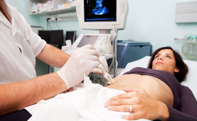
What is a biopsy?
A biopsy is a procedure where the doctor takes a sample of the suspected area to diagnose cancer. A needle is used to take the sample. The biopsy is taken with the help of a scan like an ultrasound scan or CT scan.
For patients with a suspected lung cancer, a biopsy may be taken though a procedure called bronchoscopy. For suspected cancer in the stomach and intestine, endoscopic biopsies are taken to confirm the diagnosis.
Types of biopsies
Incisional biopsy – This is a type of biopsy where a part of the cancer is cut out
Excisional biopsy –Here the entire tumour is removed with a margin of normal tissue around it
Core biopsy – This is a common method of biopsy where a core of tissue is removed to confirm the diagnosis
Fine needle aspiration cytology (FNAC) – In this type a thin needle is used to take a sample of a few cells.
Endoscopic biopsy –Here, an instrument such as a bronchoscope, endoscope or a colonoscope is used to help in getting a biopsy.
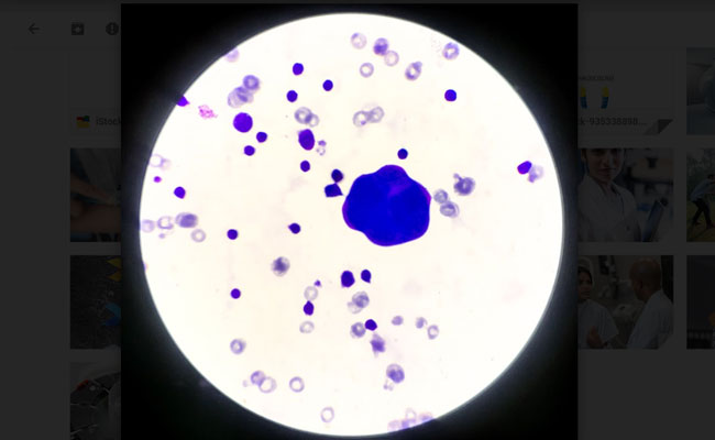
What happens after a biopsy is taken?
After a biopsy is taken, the sample of tissue is sent to the pathologist. The sample is embedded into a wax block. Then, thin sections are taken from the wax block and put on a glass slide. The slides are prepared and viewed under the microscope. Further special tests are done on the slides called immunohistochemistry (IHC). These tests will help to accurately diagnose the cancer and the type of cancer.
For some cancers, molecular testing or genetic testing is done on the biopsy sample to look for genetic changes in the cancers. Based on the results of these tests, specific types of treatment can be given to the patients. Normally about 3-7 days are needed for a biopsy to be reported, with longer time if special tests are done.
Will biopsy make cancer grow faster?
A biopsy will not make the cancer grow faster as is believed and mis communicated by some sections of the public. A biopsy is essential to make a diagnosis and then to treat cancer. No Oncologist would offer any treatment, particularly chemotherapy or radiotherapy without a biopsy result confirming the presence of a cancer. In some instances, surgery may be carried out without a biopsy if the scans suggest high possibility of cancer.
Is a biopsy procedure painful?
When a biopsy is planned, the doctor will explain the procedure and usually a local anaesthetic is given prior to the procedure. This is given as an injection and numbs the area. Therefore, most biopsies are painless but may cause some discomfort.
Bone Marrow Examination
A bone marrow examination involves taking a sample of the marrow from the bone to look for cancer cells and other abnormalities in the bone marrow. This test is commonly done in cancers related to the blood and lymphatic system. The marrow is taken from the bones of the pelvis or sternum. A local anaesthetic drug is given to reduce the discomfort prior to the procedure. The test is done on an outpatient basis and the patient can go home after the test is done. The report will be available in a few days.
Once a diagnosis of cancer is made by a biopsy and IHC testing, molecular and genetic testing may also be done on the biopsy sample to get more information about the cancer. These tests are required in some but not all cancers. They show the changes that are present at the molecular and genetic level for that type of cancer. The extra information provided by these tests help in deciding on the most appropriate treatment for the patient. In some cancers, particularly those of the blood, tests help differentiate between the types of those cancers. The tests, in some situations, may give information as to how well a treatment will work for that cancer. The different types of tests done for specific cancers are detailed in their sections.
